
WROUM, Schi, Schi, Schi, WROUM, Schi, Schi, Schi, Jiiii, Jiiii, Jiiii, Jiiii
No, we’re not at a techno concert, but in an MRI tube. Anyone who has ever had the questionable pleasure of having done an MRI scan will know what I’m talking about. As if it wasn’t bad enough to feel like Dracula in a coffin, you’re subjected to the most boring techno music in the world. As I lay there trying not to sway to the beat, I asked myself: Does it have to be that loud?
What was nuclear magnetic resonance again?
Before we go in search of the tapping and hammering, let’s remember what MRI is. MRI stands for magnetic resonance imaging and in my post about nuclear magnetic resonance (NMR) I looked at the first two parts: magnets and resonances. If you haven’t read that article yet, I recommend reading it first. As a refresher, here is the most important information in a nutshell:
MRI images do not show the density of tissue in the body, as X-ray images do, but the density of hydrogen atoms. All atomic nuclei, including those of hydrogen, are positively charged and have a spin. Spin is a quantity of quantum mechanics and describes, metaphorically speaking, the momentum with which a particle rotates around its own axis. The combination of charge and spin means that atomic nuclei are magnetic. In a magnetic field, atomic nuclei therefore rotate like spinning tops around the magnetic field lines. The number of rotations that the atomic nucleus performs per second is called the Larmor frequency.
By bombarding the atomic nucleus gyroscopes with radio waves, we can “turn them on”. However, this only works if the frequency of the radio wave is the same as the Larmor frequency – we call this resonance. Just as a toy spinning top stops spinning at some point, the pirouetting atomic nucleus slowly spins back to its resting position. As it does so, it emits radio waves that we can measure. A lot of information is hidden in these waves: about the type of atom, the atoms in the vicinity of the nucleus – and therefore about the nature of our body.

The back and forth of the radio waves can be understood as a game of blind man’s buff. We cannot see the atomic nuclei and shout “Marco?” into the body. If atomic nuclei are there and they have the right Larmor frequency, they will respond with a “Polo!” call – and we know they are there!
Flatland vs. Spaceland
Radio waves do not harm our bodies, unlike X-rays, and therefore an MRI is harmless to us. As if that wasn’t enough, it also has the impressive advantage that MRI images are three-dimensional. An X-ray image, as we know it from bone fractures, works according to the projection method. All structures of the body are projected onto the same surface and superimposed. On the basis of a flat X-ray image, we cannot tell which bone lies over which.

Tomography, as used in MRI, is also called sectional imaging. Similar to a flip-book, a three-dimensional body is broken down into many individual sectional images. Each of these sectional images shows what the body would look like if it were cut open at that point (without any blood being shed). Together, the sectional images form the 3D model of the body, from which doctors can learn more than from a flat X-ray image.
However, breaking down the body into layered images is not so easy. If you bombard a part of the body with radio waves (and shout “Marco?”), all the atomic nuclei shout “POLO!” back in chorus. From this we learn absolutely nothing about the three-dimensional structure of the body – we again only get a projection image. So how can the voices of the atomic nuclei be separated from each other?
Roses are (1,0,0), violets are (0,0,1)
We need a way to distinguish the atomic nuclei from each other. One illustrative method would be to label the atomic nuclei with different colours. We can do this neatly and systematically in three dimensions using the RGB colour space.
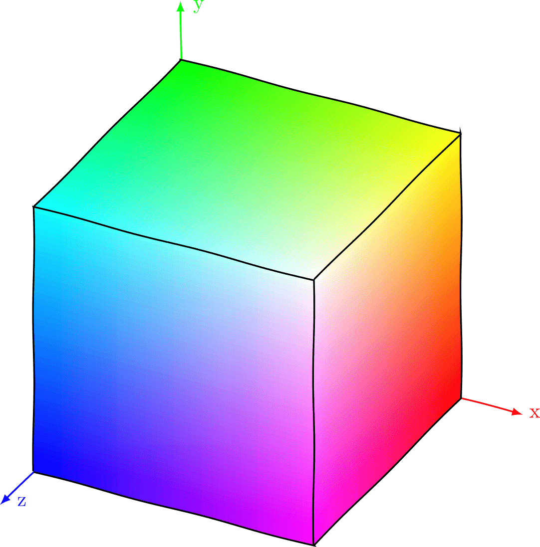
RGB stands for the three primary colours red, green and blue. Each colour can be mixed from these three primary colours. We can therefore represent all possible colours in a colour cube. The x, y and z axes denote the proportion of the three colours red, green and blue. In this way, each colour can be represented using three coordinates (Red, Green, Blue). The red colour has the easy coordinates (1,0,0) and is located at the bottom right-hand corner of the cube. Pink is the mixture of blue and red and therefore has the coordinates (1,0,1).
Red, Green, Blue are all my clothes
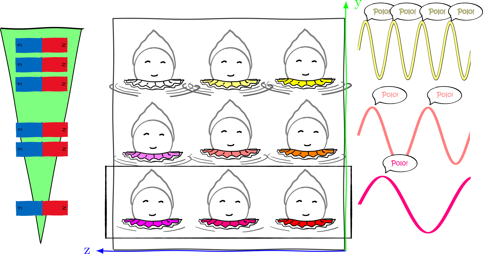
We now use the RGB principle to differentiate between atomic nuclei. Let’s take a cube of 3x3x3 pirouetting ballerina nuclei as an example. We colour the skirts of the atomic nuclei according to their position in the RGB cube and can thus clearly tell them apart. If we now shout “Marco?”, all the atomic nuclei shout back in their colour. Unfortunately, we can’t put skirts on real atomic nuclei and they don’t do us the favour of communicating with colourful speech bubbles.

But in principle, the same thing happens in MR tomography: we try to resolve the x, y and z (red, green and blue) components of the cube. However, we use magnetic fields instead of colours. In the MRI tube, there is a strong magnetic field that surrounds the entire body. This is necessary to cause the atomic nuclei to spin. In addition, three weaker magnetic fields are applied, which become stronger and weaker along the three axes (of the colour cube). We call these three magnetic fields gradient fields. Just as the red component of the colour cube increases along the x-axis, the (“red”) gradient field in the MR tomography also becomes stronger along the x-axis.
By skilfully combining and switching the gradient fields on and off, the position of the atomic nuclei can be broken down precisely. This works in three steps, as I will explain in more detail below: First, a layer with a certain amount of red is picked out, then a row with a certain amount of green and finally a column with a certain amount of blue. What remains is a single pixel whose position (colour) we know exactly.
Red: Choice of layer
The first step of the slicing process is (surprise) to cut a slice out of the 3D body. We start with the x-direction. In our RGB image, this means that we define the red component of the atomic nucleus. To do this, we create a gradient field in the x-direction, i.e. the magnetic field becomes stronger in the x-direction. This changes the Larmor frequency of the atomic nuclei – the “redder” atomic nuclei rotate with greater momentum. If we select a “particularly red” Marco pulse, it will only turn on the fast-spinning atomic nuclei with a high red component. Only the atoms in this layer answer us with their Polo-call, all others remain silent. As a side note: In reality, the radio wave is not actually red, but its frequency is equal to the Larmor frequency of the atomic nuclei in a particular layer.

Green: Selection of the line
We no longer have an atomic cube, but only a flat disc. All the atomic nuclei in this disc are spinning and will shout “Polo” whenever they look at us. In order to tell the rows – i.e. the green part – of these atomic nuclei apart, we apply another gradient field, this time in the y direction, after we have emitted the Marco pulse. This changes the Larmor frequency of the atomic nuclei. Or to put it more simply: the atomic nuclei that are in a stronger magnetic field rotate faster than the nuclei in the weaker field. The nuclei in the top row will therefore shout “Polo” more often than the nuclei in the bottom row. The signal emitted by the atomic nuclei is recorded by a computer and broken down into different frequency components so that we can distinguish between the rows.
Blue: Selection of the column
Selecting the column, i.e. determining the proportion of blue, works in a very similar way. However, we cannot use exactly the same trick. The blue and green gradient fields would simply overlap and we would no longer be able to determine the exact position. Instead, we switch on the blue gradient field in the z-direction only very briefly. As a result, the atomic nuclei in the left-hand column only rotate faster for a very short time, giving them a head start over the nuclei in the right-hand column. The point at which the atomic nuclei look at us and shout “Polo” is therefore slightly offset. We call this phase shift. We can calculate this information from the signal with the help of computers and thus assign the columns correctly.

26 M&Ms made from pure gold
The procedure just described gives us information about one pixel of the image. In fact, not even that, as several passes are required to analyse the phase information. An MRI image consists of thousands of pixels and therefore it takes much longer to produce an MRI image than an X-ray image (15-30 minutes compared to an instant). And not only does it take longer, it is also ten or even a hundred times more expensive than an X-ray, which can be obtained for the bargain price of €10 to €50.
The reason for the high costs is the strong magnetic field that prevails in the MRI tube. To get a good signal, the Larmor frequency must be high and the magnetic field strong. Fields with a strength of 1.5 to 3 Tesla (not related to the car) are common – hundreds of thousands of times stronger than the earth’s magnetic field. Superconductors are required to generate such strong magnetic fields, which only work at near absolute zero (-273°C). Due to the extreme cooling temperature, the coolant, liquid helium, is correspondingly expensive. Operating an MR tomograph costs a good 15,500 euros per year – you could buy about 26 M&Ms made of pure gold for that.
Techno from the tube
The gradient fields, on the other hand, are around a hundred times weaker than the global magnetic field and are generated by current-carrying coils. Several hundred amperes of current are chased through the coils (a mobile phone charges with around 2 amperes). Due to the prevailing magnetic field, the charge flowing through the coils is subjected to an enormous force that is 120 times stronger than the force of gravity. The coils are torn back and forth every time the current is switched on and off. They start to vibrate and this is the cause of the loud hammering and whirring in the MRI tube. The volume can be as loud as that of a roaring underground train or a rock concert.

Excessive ticket prices, bad music, earplugs to block out the noise, little freedom of movement and the occasional use of tranquillisers – an MRI walk has more in common with a techno concert than originally thought. And if you imagine atomic nuclei as ballerinas, there are also wild dancers and lots of bright colours in both cases.
In this article, I explained MRI, which is one of the first-generation quantum technologies. You may also be interested in the article on the first quantum revolution. Do you like what you read? Then subscribe to my blog and never miss a new post again!